|
The ciliary muscle, part of the uvea, passes from the deepest layer in the the cornea to the anterior region of the sclera. In mammals there are two muscles, Bruke and Muller. In birds, there is an extra Crampton muscle. The contraction of these muscles raise the pressure in the vitreous. This pushes on the lens, which is being held peripherally by the iris, shaping it to allow light to hit the retina. In humans the opposite happens. The ciliary muscles act on the lens to release pressure, letting the elasticity determine its shape. The ciliary muscle and the iris are smooth in mammals, but striated in the avian eye.
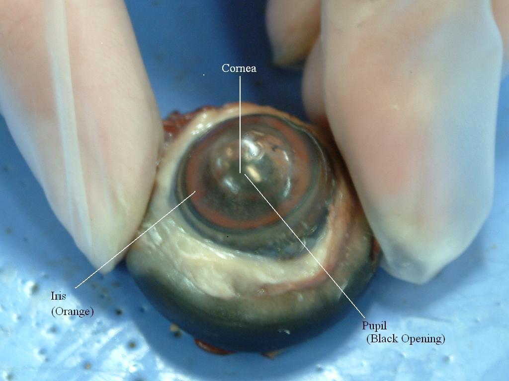
All vertebrate eyes contain an iris, which controls the size of the pupil. In birds, the color of the iris depends on the behavior of the particular species. The singing varieties contain brown irises, while birds of prey have yellow irises. Usually birds have a black edge around the iris, which makes the pupil appear larger.
The pupil is round in avian and mammalian eyes. The only exception is the owl, who contains an oval pupil with a long horizontal axis.
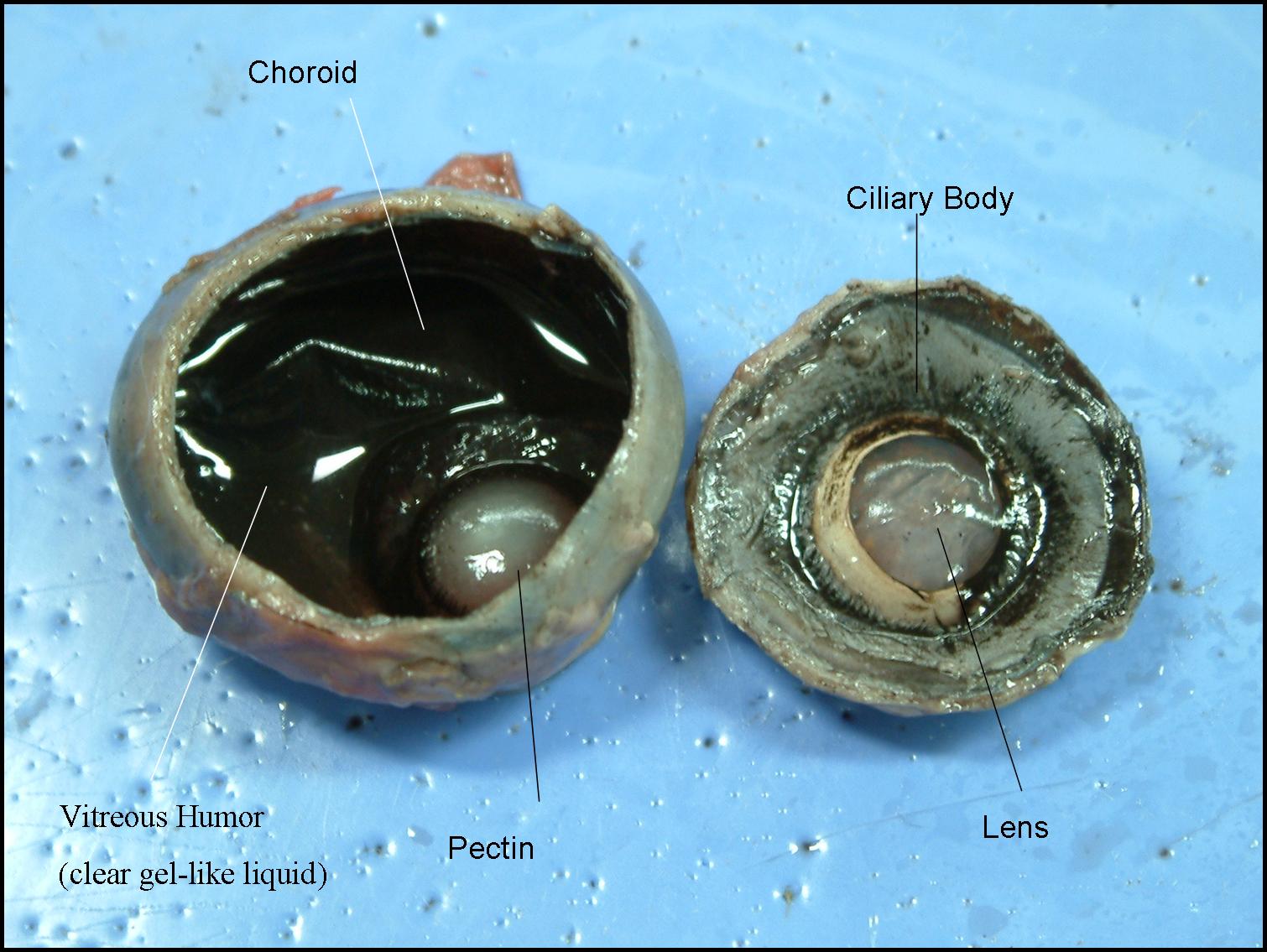
Pecten is a structure found in birds but not in mammals. It is vascularized, supplying the bird with nutrients and oxygen for the eye and carrying away waste. It is composed of connective tissue with many folds and is highly pigmented, extending from the optic disc into the vitreous. In some birds, such as swans and ducks, it extends to the lens. Pectin is an ectodermal structure and homologous in the cone of reptiles. Some think pecten has erectile properties, which is a defensive mechanism against too strong light. Others think that pecten can regulate tension, secretion, and temperature at high altitudes.
Birds, like mammals have aqueous
humor and vitreous humor.
Aqueous humor is located between the cornea and lens. Vitreous humor
is located between the retina and lens. In both organisms they have
the same function.
 |