|
In embryology, the optic placode gives rise to the optic vesicle. This is where the eye begins its growth. After development, the optic vesicle continues its connection with the brain and is termed the optic stalk. This is what carries the information to the brain, which allows the images received by the eye to be interpreted. The optic stalk is an extension of the brain, but is referred to as the optic nerve, which is the second cranial nerve.
Although the retina responds to light intensity and wavelength, the brain is what actually gives the image contour, orientation, and motion. The visual information is consciously received in the brain, which also merges the images seen by the two eyes. The fibers in the optic nerve also send information that allows motor output of the eye muscles, as well as muscles within the neck and trunk to turn toward the visual stimulation. Interpretation of visual input starts in the retina (as much as 90% actually occurs there) and continues as it travels through the optic nerve into the brain. This process is the same in both the avian and mammalian classes.
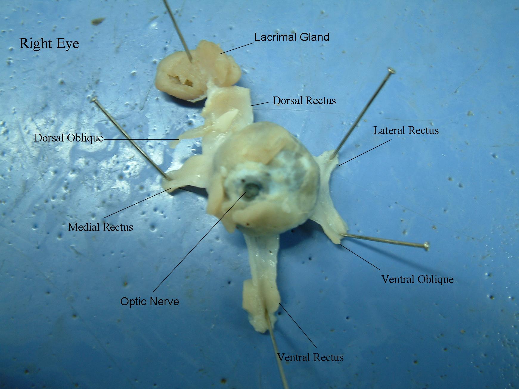
The lacrimal gland is what supplies tears, which moisten
the eye and aids in the immune system. In most animals there are two canals
that connect the gland to the eye, although in the pig, rabbit, and ewe
there is only one passage. It lies within the fossa of the frontal bone
in humans, and the gland does not function fully until after three months
of age.